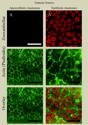
Farah Hussain: Does the cytoskeleton of the sea anemone, Aiptasia pallida, change as a result of its symbiosis with zooxanthellae?
Differences in the actin cytoskeleton between aposymbiotic (left column, no Symbiodinium) and symbiotic anemones (right column). Autofluorescent Symbiodinium, red; filamentous actin, green. Farah Hussain '09
Corals are integral to many worldwide marine ecosystems but are threatened by bleaching, a process in which stress results in the loss of symbiotic zooxanthellae. To better understand the symbiont-host relationship, we studied the cytoskeleton of a related cnidarian, the sea anemone
Aiptasia pallida in the presence and absence of zooxanthellae. We observed that zooxanthellae densely populated host gastrodermal cells, occupying the majority of intracellular space. This suggests that anemones must manipulate their cell shape and cytoskeleton in order to perform normal functions while accommodating symbionts. By staining cortical F-actin with a small-molecule dye, we observed that host cells harboring intracellular zooxanthellae were larger in cross-sectional area than aposymbiotic cells. There were also striking differences in the shape of these cells. Symbiotic gastrodermal cells exhibited compact curves that fit snugly over the intracellular symbionts. In contrast, aposymbiotic cells were smaller and polygonal. These observations indicate that the host rearranges its cytoskeleton and thus changes shape to accommodate symbionts. Microtubules visualized by immunofluorescence showed putative cilia on the surface of tentacles. In the ectoderm, staining showed potential outlines of nuclei, nematocyst capsules, and the outlines of vacuoles. Staining in the gastroderm was not reproducibly detectable, due most likely to problems with permeabilization. This investigation suggests that there are changes in cell shape and size as a result of the symbiosis between Aiptasia and zooxanthellae.
Ian Yarett: The spatial distribution of proliferating cells during tentacle regeneration in the sea anemone Aiptasia pallida.
EdU indices in the ectoderm at various regions along the length of each intact (red; n=9) and regenerating tentacle (blue; n=9). The proportion of cells undergoing DNA synthesis in both intact and regenerating tentacles drops sharply from the proximal to distal regions of the tentacle. Ian Yarett '09
Anthozoans, along with many other cnidarians, are able to rapidly regenerate after undergoing tissue damage. In this study, I analyzed the spatial distribution of proliferating cells in intact and regenerating tentacles of the symbiotic anemone Aiptasia pallida. The percentage of cells in S phase at various regions along each tentacle was directly measured by incorporation and visualization of the thymidine analog 5-ethynyl-2'-deoxyuridine (EdU), allowing an indirect assessment of cell proliferation in vivo. A proximal-distal gradient of cell proliferation was observed in both intact and regenerating tentacles, with the most DNA synthesis at the base and the lowest at the tip. Furthermore, the process of regeneration did not lead to an increase in the levels of DNA synthesis compared to that of intact tentacles. The results suggest that Aiptasia grow in a manner similar to hydra, in which the site of cell division can be spatially decoupled from the region undergoing growth or tissue repatterning.
Julia Berthet: The spatial distribution of proliferating cells in the sea anemone Aiptasia pallida during different states of symbiosis.
Confocal image of a tentacle of the symbiotic anemone Aiptasia pallida. Zooxanthellae autofluoresce red in the anemone's gastrodermal cells. Replicating nuclei are labeled with EdU (cyan) and all nuclei are stained with TOTO (green). Julia Berthet ‘10
During growth and recovery from bleaching cnidarian hosts must acquire new symbiotic dinoflagellates so as to meet metabolic demands for photosynthetically fixed carbon. In order to better understand the dynamics of cell division and symbiont propagation within new tissues, I characterized the distribution of cells in S-phase, detected in vivo through the incorporation of the nucleotide analog 5-ethynyl-2'-deoxyuridine (EdU). Indices were examined in the ectoderm and gastroderm of Aiptasia pallida in three different symbiotic states: Aposymbiotic, repopulating, and fully symbiotic. Upon exposure to symbionts, levels of proliferation increase dramatically and Aiptasia pallida appear to modulate the distribution of proliferating cells, transitioning from an even distribution of ectodermal growth to a decreasing gradient of proliferation along the tentacle. Aiptasia undergoing repopulation therefore display patterns of cell proliferation, which closely resemble the fully symbiotic state, despite wide disparities in the density of endogenous symbionts, suggesting that symbiont exposure initiates new patterns of cell cycling independently of nutrient availability. Gastrodermal proliferation alternatively exhibits declining proximal-distal gradients of proliferation in aposymbiotic and symbiotic states. During repopulation anemones exhibits a uniform distribution of cells in S-phase, indicating a role for host proliferation in symbiont distribution.
Interdisciplinary Special Majors
Swarthmore students may pursue interdisciplinary curricula developed by the faculty, or they may take the initiative to define their own distinct academic program with the support of faculty mentors.