Regulative
Development in Axolotl Embryos; Splitting the Heart
Field

|
|
|

Results
Six axolotl embryos were initially prepared successfully, and of those, at least one showed a distinct heart beat by 6 days after surgery. Determining whether or not the heart field had been affected by the graft was difficult due to the opacity of the axolotl skin, and the positioning of the embryo in the operating dish; one side down, such that only one side could be viewed at a time without damaging the embryo.
Evidence that the heart field was not particularly affected includes the fact that the grafted tissue was at least partially rejected. Additionally, when viewing the gills close up, the flow through them seemed completely synchronous between the left and right gills, as well as in synch with the visible heart beat. As for the stage of heart development, it appears to have developed at least somewhat normally to a point somewhere between stages A and B in Figure 7. Specifically the blood flow seemed to start toward the ventral-anterior corner of the heart, flow upwards toward the dorsal area, and finally down to the ventral-posterior region (Figures 5 and 6).
There were some irregularities however. In Figures 3, 4, 5 & 6 we can see that the heart region is well behind the gills. This is somewhat farther back and more to one side than one would normally expect to find the heart. This irregularity opens up the possibility that the heart field was at least somewhat affected, despite appearing to have a synchronous heart beat.
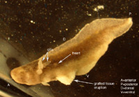
Figure 3. Photo of six day axolotl embryo showing location of heart and early heart development. Note eruption of previously grafted tissue from ventral region.
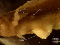
Figure 4. Close up of figure 3, focusing on heart region and tissue eruption.
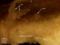
Figure 5. Close up of figure 3&4, focusing on heart region.
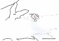
Figure 6. Outline diagram of figure 5, showing specifically the outline of the heart region. The red arrows indicate the observed direction of blood flow.
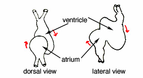
A
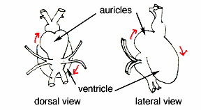
B
Figure 7. Two early stages of amphibian heart development; heart development in above figures 3-6 is presumed to be between these two stages. Diagrams simplified after Rugh, 1951.