Regulative
Development in Axolotl Embryos; Splitting the Heart
Field
|
|
|

Discussion
From our observation of a single distinct heartbeat, it appeared that the attempt to divide the heart field was not successful. Additionally the observed direction of blood flow and apparent synchronous flow through the left and right gills seemed consistent with normal amphibian heart development (Rugh, 1951). However, the ability to observe the heart beats was hindered by the opacity of the axolotl skin, as well as the position of the embryo in the operating dish, so these observations may not accurately reflect the true condition of the heart. In addition, the heart appeared to be somewhat more posterior than would normally be expected. The presence of this irregularity leads us to believe that the heart field was affected in some way. If this were the case, the heat could actually be split, or partially split, but have two heart beats that happen to appear synchronous.
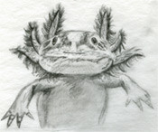
The fact that the grafted tissue was observed erupting from
the belly/ventral region of the embryo may suggest that the
graft, which was intended to split the heart region, was not
incorporated into the embryo enough to sufficiently block
communication between the two halves of the heart field
(Figures 3, 4,&5).
It should be noted that there are other explanations for
these data that might also be evidence for regulative
development. For instance, the gill tissue could be
transforming into heart tissue through cell-cell
interactions. However, the relatively advanced developmental
stage of the donor tissue, and the fact that the photos seem
to clearly show tissue rejection, do not support this
conclusion. In the end, these results are at best
inconclusive in regards to regulative development in the
formation of the axolotl heart.
It should be noted however, that previous experiments using
small pieces of sterile foil as a barrier rather than gill
tissue, did yield results supporting the existence of
regulative development in the axolotl heart. Specifically,
two hearts, one on either side of the foil barrier, were
seen to develop and clearly beat asynchronously (Vélez
and Krsmanovic, 2004). Other examples of the
manipulation of morphogenetic fields, and therefore evidence
of regulative development, have been shown in limb
regeneration studies. Axolotls are actually able to
compensate for damage to the limb morphogenetic fields
throughout their lives, to the point where entire limbs may
be regenerated from the remaining tissue of a limb stump
(Gardiner et al 2002).
Since studies show that there is ample evidence for
regulative development in axolotls it would therefore seem
that the our results likely have more to do with
experimental technique than absence of regulative
development in axolotls. While this tissue graft procedure
was not particularly complex, and was the method originally
suggested in Hamburger's Manual of Experimental
Embryology, in retrospect it does not seem well suited
to those who have had little to no practice doing
microsurgery. Additionally, it should be noted that while
the embryos were largely at the appropriate stage at the
beginning of the procedure, as time went on, the warmth of
the room allowed them to develop more quickly than we had
anticipated, leading to fewer suitable embryos. It is
important to remember that there is only so much time before
the heart primordia fuse, and grafts must be done
before this happens in order to be sucessful. In
the future it would be advisable to keep the embryos cold
whenever they are not actually being worked on, rather than
letting a number of them sit out at room temperature.