|
Procedure:
1. Obtain axolotl embryos. For this experiment, we
will be using wild type and albino axolotl embryos at stage
21 in their staging series.
2. Prepare the axolotl embryos as follows:
a. manually dejelly embryo of appropriate stage and keep in
HEPES-buffered
Modified Steinberg's solution (HBSt)
containing antibiotics until use. For microsurgery, also
remove membrane around embryo.
b. place embryos into operating dish containing 1 x HBSt and
antibiotics
c. use clean technique; dip tools, pipettes, and glass
bridges in 70% EtOH and then into sterile HBSt before
using
3. Start microsurgery
on your donor embryo. Locate the gill organ field which is
in the first anterior third of the embryo (toward head) and
right above the small crest or indention closer to the
ventral side (Rugh, 1962) . Isolate the gill organ field (or
the region that will become the gill organ field) by cutting
around it and detaching that specific piece of tissue from
the embryo.
4. Start transplantation on your recipient embryo. Cut a
small slit in a region close to the gill organ field on the
recipient. Gently slip donor gill organ field into slit and
be sure to secure the graft in place. It is critical to try
to get the graft as far under the cut as possible to ensure
that the recipient will begin to incorporate the new tissue
into its development process.
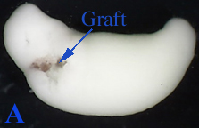 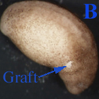
Figure 1:
A. A 21 stage albino axolotl embryo
with a transplanted wild type axolotl graft (supposedly the
gill field). The picture was taken immediately after
microsurgery.
B. A 21 stage wild type axolotl
embryo with a transplanted albino axolotl graft. The picture
was taken immediately after microsurgery
|