|
|
|
|
|
|
|
|
|
|||
|
|
Results |
|
||
|
|
|
All four groups of embryos were observed under a fluorescent microscope. The trunk region was observed for areas of brighter cells. In picture A, vertical lines of cells are slightly brighter than surrounding cells. This is where the motor neurons will develop. In picture C, we see evidence of a row of somites developing along the trunk region. The controls show less evidence of developing motor neurons or somites. Figure 2. Pictures of fixed zebrafish embryos under a fluorescent microscope 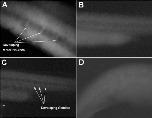 A-Trunk region of a 24 hour zebrafish embryo treated with motor neuron specific ZNP-1 antibodies B-Trunk region of a control, unstained 24 hour zebrafish embryo C-Trunk region of a 24 hour zebrafish embryo treated with somite boundary specific F6 antibodies D-Trunk region of a control, unstained 24 hour zebrafish embryo |
||
Last Modified: 2 August 2001
[Lab
Protocols
| Students
| Cebra-Thomas
| Course
| Links
]