|
In the sea urchin, early cell
divisions are rapid; the cell cycle alternates between S
phase, where new DNA is synthesized, and mitosis. As a
consequence, the embryonic DNA is not transcriptionally
active; it is not actively "read" to produce new messenger
RNAs (mRNAs). The proteins that are synthesized during
cleavage utilize mRNAs in the cytoplasm provided by the
mother (Fig. 7.30).
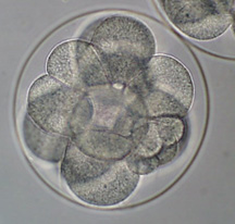
The
first 3 cell divisions bisect the embryo equally (Fig. 8.8,
staging
series). The first 2 cleavage
planes from the "top" (known as the animal pole) to the
"bottom" (known as the vegetal pole), while the third runs
across the equator and separates the embryo into "animal"
and "vegetal" halves.
The 4th
cleavage is more unusual. The cells in the top half divide
equally, but those in the bottom half divide unequally,
creating large cells (macromeres) and small cells
(micromeres). This is accomplished by one of the centrioles
positioning itself in the middle of the cell, so that the
spindle is displaced to one side (Fig. 8.10).
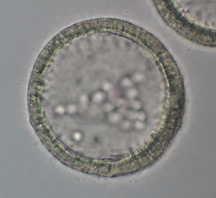
The
cells continue to divide until they form a hollow ball known
as the blastula (Fig. 8.11). Each of the cells produces a
cilia. At this point, the genome is activated and starts to
express new genes. One of these genes codes for a protease
that digests a hole in the fertilization envelope; the
embryo "hatches" and begins to swim.
Shortly
after hatching, the descendants of the micromeres at the
vegetal end detach from the epithelial sheet and move into
the blastocoel (ingression). These are known as primary
mesenchyme cells and they form the calcium carbonate
spiculesof the larval
skeleton (Fig. 8.18, 8.19, 8.20).
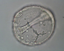
The
decendants of the macromeres thicken to form the vegetal
plate, which invaginates
to form the
archenteronor gut
(Fig. 8.17, 8.21). This process is
known as gastrulation and, in
addition to forming the gut, it results in a multilayered
body plan (Fig. 8.16). The archenteron extends by cell
rearrangement (Fig. 8.23) and by connections between the
cells at the archenteron tip (secondary mesenchyme cells)
and the extracellular matrix lining the blastocoel (Fig.
8.25). Once the archenteron reaches the other side, the
mouth is formed.
Click here to see movie.
As the skeleton is laid down, the embryo's shape changes to
form the prism and then pluteus
larvae(Fig. 8.17).
|