|
|
|
|
|
|
|
|
|
Staining Drosophila melanogaster imaginal discs for engrailed gene expression using the ß-galactosidase reporter
Carballo L, Hwang B Swarthmore College, Swarthmore, PA BIO 024, Cebra-Thomas
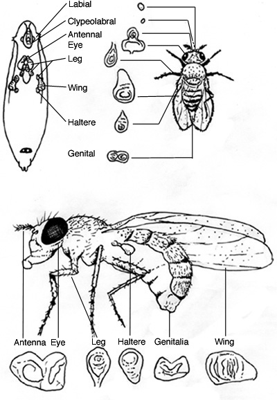
Introduction: Drosophila melanogaster, commonly known as the fruit fly, passes through the larval and pupal stages before reaching the adult stage (Gilbert, 2003). A newly hatched Drosophila larva resembles a miniature worm and is called the first instar larva. The larva molts in 25 hours and grows into a larger form called the second instar larva. Another molting takes place after 24 hours and the larva grows into the third instar larva and starts to climb out of its food source to undergo pupation. After 30 hours, the third instar larva molts into a pupa (Tyler, 1994). It is during the pupal stage that the larva metamorphoses into the adult fly. During metamorphosis, most of the larval structures are lysed and replaced by adult structures. Programmed apoptosis destroys most of the larval body, while new adult structures develop from clusters of undifferentiated imaginal cells set aside within the larva (Gilbert, 2003). Imaginal discs are one of the main groups of cells that compose the imaginal cell population. Imaginal disc cells give rise to structures like the legs, wings, antennae, eyes, thorax, and genitals. Unlike most larval cells that go through limited cycles of divisions, imaginal discs divide rapidly (Gilbert, 2003). Moreover, imaginal disc cells begin to divide strictly during metamorphosis. The dividing imaginal disc cells undergo proliferation, differentiation, and elongation, which together constitute the process called eversion (Gilbert, 1993). The manner in which imaginal disc cells evert into their respective structures is often described as "telescoping out" due to the particular way that adult structures are folded into their respective discs. Eversion occurs without cell division as disc epithelial cells simply change their shapes (Gilbert, 2003). Although disc cells remain undifferentiated until metamorphosis, specification of their general cell fates takes place during the embryonic stage. Cell fates become increasingly specified as the larva moves though the different developmental stages. Transcription factors are responsible for specification of cell fates and axis patterning of the imaginal discs. For instance, it is known that compartmentalization and anterior-posterior patterning in the Drosophila wing discs are accomplished through complex interactions between a group of genes that includes engrailed, decapentaplegic (dpp), splat, and oculomotor blind (omb) (Gilbert, 2003). Because of its posterior localization pattern, engrailed is believed to specify posterior regions of imaginal discs. There are many methods that are used to visualize gene expression patterns in the imaginal discs. Widely employed methods include in situ hybridization, antibody staining, and the use of reporter genes. Reporter genes code for enzymes or detectable proteins and their coding regions are fused into the promoter of a gene whose expression is to be monitored. Hence, when the genes of interest are expressed, reporter genes are concomitantly expressed. Well-known reporter proteins include alkaline phosphatase, b-galactosidase, bacterial luciferase and various fluorescent proteins (Gilbert, 2003). The purpose of this experiment is to use b-galactosidase to visualize the expression pattern of engrailed in Drosophila imaginal discs. b-galactosidase is an enzyme encoded by lacZ of the E. coli lac operon. X-gal (5-bromo-4-chloro-indolyl- b-D-galactoside) will be used to analyze tissue specific expression of b-galactosidase. b-galactosidase cleaves galactoside from X-gal, a chromogenic substrate, to yield 5-bromo-4-chloro-indolyl. 5-bromo-4-chloro-indolyl then is oxidized into 5-bromo-4-chloro-indigo, which is colored (Cruz, 1993). If the promoter lacZ is successfully incorporated into the Drosophila genome such that it is under the control of regulatory elements of engrailed, then imaginal discs of engrailed-lacZ mutant Drosophila should stain according to the known engrailed expression pattern.
Figure. 1. Cruz. Figure.17.2. Location and identification of Drosophila imaginal discs
Objective: The objective of the experiment is to determine the expression pattern of the engrailed gene in Drosophila melanogaster imaginal discs using the reporter ß -galactosidase.
|
||
|
|