

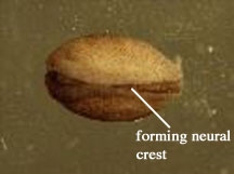
Figure 1. A pigmented stage fourteen embryo. Stage fourteen embryos are characterized by the closing of the neural tube. The name "keyhole stage" has been used for stage fourteen due to the unique shape of the neural crest. Pigmented embryos such as this one were used as recipient embryos in the transplant procedure.
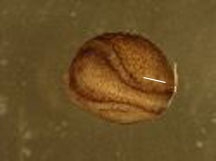
b)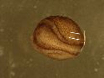
c)
Figure 2. A step-by-step schematic of the incisions necessary to excise a region of the axolotl embryo. a) This initial incision should be made on both the recipient embryo (to become the site of tranplantation) and the donor embryo. b) The second parallel incision to be made alongside the initial incision of the donor embryo. c) These two small cuts should be made perpendicular to the first two incisions in the donor embryo in order to free a region of the neural fold for excision and transplantation.
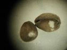
Figure 3. Sucessful transplants of regions (circa twenty-four hours after transplant) near the neural crest region of the axolotl embryo. The transplanted region orginating from an albino embryo can be easily identifiedin the recipient pigmented embryo as a region devoid of pigment.
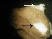
Figure 4. Developmental abnormalities leading to the termination of development. The transplanted pigment-free region (indicated by the black arrow) had begun to heal before development failed. However other regions of the embryo failed to develop properly (indicated by the white arrow).
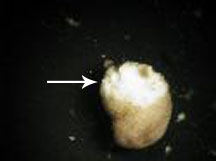
Figure 5. A failed transplant of the neural crest region in an axolotl embryo. The cells of the graft failed to adhere to the surrounding cells in the recipient embryo, possibly because of incorrect alignment of the ectodermal and endodermal cells of the donor and recipient. Development arrested less than forty eight hours after the surgery.