|
|
|
|
|
|
|
|
||
|
|
|
Grafting Procedure: Of the almost one hundred embryos used in the grafting procedure alone, only sixty-two survived the dejellying process. Twenty-one embryos were killed during the demembranating procedures. Twenty were in the early neurula II or III stage (stages 15 or 16) of development and appropriate for this experiment (Bordzilovskaya, 210). Eleven embryos were killed during the grafting surgery. These embryos generally had too large an incision made down the midline of the ventral side, which resulted in the spilling out of the light tan colored endo- and mesodermal cells (Figure 3). Only nine embryos were successfully grafted and set aside with the two control embryos Forty-eight hours after surgery, most of the embryos had reached stage 27 and some of the embryos were moving by themselves (Figure 4). One embryo responded to touch by wiggling the anterior half its body back and forth. Four of the embryos had damage near the anterior end, consisting of an area where the dark ectodermal cells were missing or barely attached to the body and the lighter colored endo- and mesodermal cells were visible or hanging out the body (Figure 5A). Clumps of cells had fallen out of the embryos and were floating in the HBSt solution. After five days, seven of the nine test embryos had disintegrated into an unorganized mass of cells (Figure 5B). By day six, all embryos, including the controls, had died.
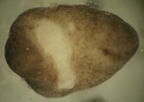 Figure 3. Surgically damaged recipient embryo. The incision was too large, resulting in the loss of light tan colored endo- and mesodermal cells. Embryos damaged in this way were ineligible for the experiment.
 Ne Ne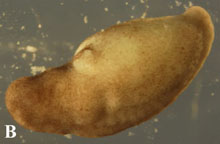
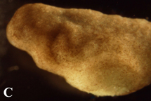
Figure 4. Post-operational development of test embryos. (A) 12 hours after operation, graft is still clearly discernable from host embryo tissue. (B) 48 hours after surgery, embryos had grown to stage 27. The graft is partially covered with host ectoderm cells and is becoming less prominent. (C) By 72 post-surgery, embryos had developed to stage 32 and the incision was barely visible.
 Need Need Figure 5. (A) Injured stage 34 embryo four days post surgery. Embryos were found with injuries near the anterior head-forming region and often had lost some endo- or mesodermal cells as well. (B) Embryos six days post surgery. Embryos eventually disintegrated into masses of cells with no discernable organization. |
![]()
![]()
![]()
Last Modified: May 13 2004
[Lab
Protocols
| Students
| Cebra-Thomas
| Course
| Links
]