

Figures
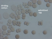
Figure 1. Experimental group one hour after first twinning event. Single-cells in this image indicate a failure to fertilize. The fertilized embryos are at varying stages of development: some are at the four celled stage, while some are still dividing to the two celled stage. Once the majority of the embryos has reached the four celled stage, the sample was twinned.
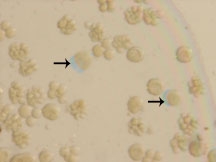
Figure 2. Single division experimental group twenty-four hours after fertilization. Note the bluish shadows associated with some of the embryos. The shadow indicates movement, and therefore the presence of motile pluteus larvae.
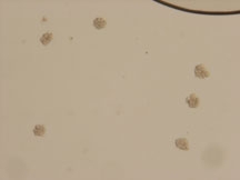
Figure 3. An experimental group twenty-four hours after second twinning event. Note that no embryos have developed fully, though partial development has continued after the twinning events.