Feather
Bud Development
Discussion:
In both experimental groups, the skin patches grew as expected from the initial placode stage producing feather buds (figure1); however, separation from the embryo retarded the development of all of the cultures. That is, whereas chick embryos at approximately 22 days (our oldest culture) would have been developing feather filaments, our cultures arrested at late long bud stage, right before feather filaments could form (figure 5). The more developed feather buds did allow us to determine what would have originally been the anterior and posterior of the chick; as the feather buds develop, especially along the spinal tract, they tend to lean toward the posterior of the chick (Chuong and Widelitz, 1998).
In addition to the skin, our limb cultures also showed feather bud development. Although the appendages did not show any bone or muscle etc. development, the epidermal layer in contact with the media formed feather buds similar to what was observed in our other cultures (figure 7).
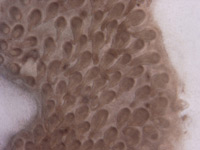
The irregularities we observed in some of the cultures (skin clumped together; no specific pterylae patterns; irregularly shaped dermal papilla) were likely the result of these segments being cultured on the underlying membrane without the mesenchymal side facing the media, or not lying flat on the culture insert (figure 6). Either of these cases would cause abnormal, clustered growth. In addition, it is also possible that the positioning of the skin on the culture inserts affected the paths of the promoter and inhibitor signals (functioning between the epithelium and mesenchyme) that help to form chick feather buds. In recent studies, both fibroblast growth factors and bone morphogenetic proteins (FGF, BMP) have been found to influence the pattern of feather bud development; their interactions determine the feather bud tracts (Rouzankina et al., 2004). If these signal pathways, or their respective gradients, were to be altered, deformed feather buds and tracts would be produced.
In addition to some irregular feather bud growth, it was noted that all skin cultures stopped growth at approximately the same point, when feather buds started to elongate and resemble small pieces of string (figure 5). This demonstrates that the feather buds ceased growing before gaining more of their "feather" character at the feather follicle/ feather filament stage (figure 2C). Growth ceased even though the culture media was replaced, indicating that perhaps this method of culture does not provide the necessary nutrients or structural support for the skin to develop past a certain stage. Perhaps this is similar to the results of shell-less chick cultures, where embryos do not produce complete skeletons since calcium (needed for bone development) cannot be derived from the shell (Cebra-Thomas, 2004).
Building upon the results of this and previous experiments, it would be interesting to investigate the cause of the arrested growth of skin in culture. Skin could be removed at various stages of embryonic maturity to determine at what stage in development the ability to culture stops. The results of such an experiment could have implications in medical research and the development of skin grafts for burn patients, or for artificial skin used to treat diabetic ulcers and other ailments (Organogenesis, 2003). If the cause of stunted growth in skin cultures could be determined, perhaps grafts could be produced on a larger scale and expenses lowered.