|
|
|
|
|||||||||||||||||||
|
|
|
||||||||||||||||||||
|
|
Figures Concentration # Embryos Alive # Embryos Dead Control (ZES) 16 2 0.025 M 21 8 0.05 M 16 13 0.1 M 16 17 0.2 M 0 24 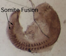 Figure 3. F6 antibody stain for somites
in an embryo treated with 0.05 M valproic acid solution.
Somite fusion of adjacent pairs and mis-segmentation of
somites are characteristic of Class I anomalies. 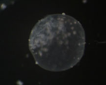 Figure 5. Zebrafish embryo treated with
0.2M valproic acid solution that failed to develop. |
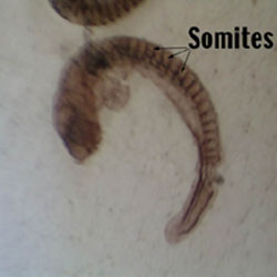 Figure 2. F6
antibody stain for somites in control embryo demonstrating
normal somite development at 28 hours after
fertilization. 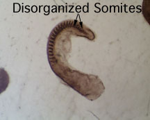 Figure 4. F6 antibody stain for somites
in an embryo treated with 0.1 M valproic acid solution.
Disorganized and scrambled somites are characteristic of
Class II anomalies. |
Last Modified: 31 May 2001
[Lab
Protocols
| Students
| Cebra-Thomas
| Course
| Links
]
![]()
![]()
![]()
