|
|
|
|
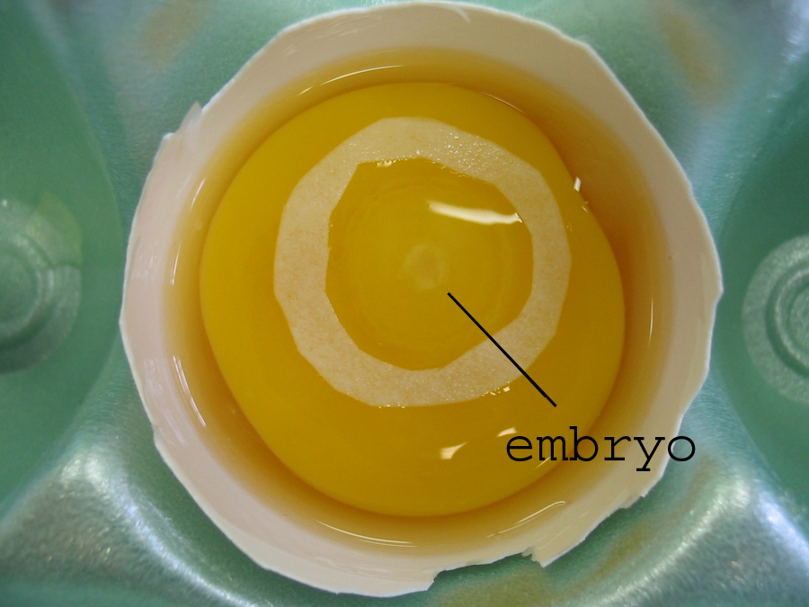
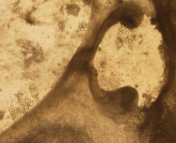
Figure
1. First method of transferring the embryo
directly from the shell to the culture dish; here,
an opened shell with the embryo exposed, surrounded
by a filter paper ring.
|
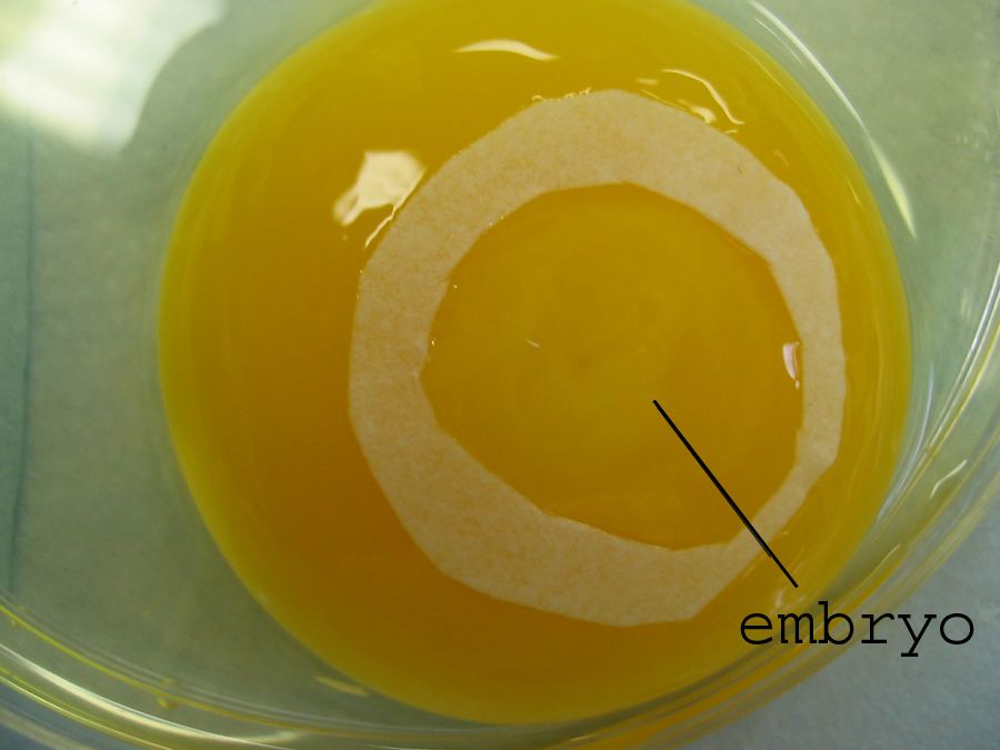
Figure
2. Second method of transfer; here, the
intact yolk has been moved to a petri dish and the
embryo isolated with a filter paper ring.
|
|
|
|
|
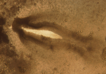
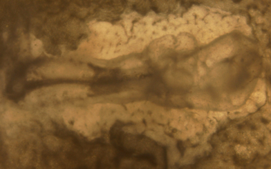
Figure
3. A stage 8 chick showing dark shadowy
regions that indicate the bilateral heart primordia
on either side of the notochord.
|
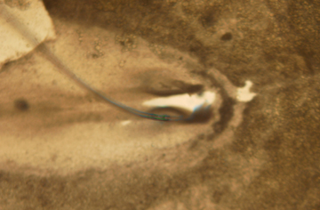
Figure
4. A glass taper (extending from top left
corner) can be seen enlarging the surgical incision
made in the anterior portion of the chick
embryo.
|
|
|
|
|
Figure
5. A glass taper (extending from top left
corner) can be seen enlarging the surgical incision
made in the anterior portion of the chick
embryo.
|
Figure
6. Surgery gone wrong: a two-headed
embryo, presumably caused by an incision that
bisected too much anterior tissue.
|
|
|
|
|
Figure
7. An embryo with two beating hearts, one
on each side of the notochord.
|
Figure
8. This chicken embryo has an abnormal
heart, probably due to late fusion of more fully
developed heart primordia.
|
|
|
|
|
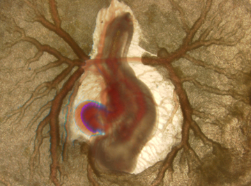
Figure
9. A stage 21 control chick with a
functioning circulatory system – the heart is
beating, which is noted in this photograph as a
blue outline surrounding the moving tissue.
|
Figure
10. The steadily beating heart of a stage
23 control chicken embryo. The branching of the
vascular system is more complex.
|