![]()
Gastrulation in Clay
(Zebrafish)!
Anisha Chandra '06 and Lauren Fety '06, Swarthmore College, February 2004
Introduction
The zebrafish undergoes a very interesting gastrulation process. After fertilization, all the yolk streams to the vegetal pole via microtubules, leaving a non-yolky animal pole. The large amount of yolk restricts and defines this process. However, there are similarities between the gastrulation of zebrafish and other vertebrates, such as amphibians. One striking dissimilarity, though, is that in zebrafish, the entire embryo forms to one side of the overall mass, due to the size of the yolk. Another difference is that early zebrafish cells are much more loosely associated than amphibian cells and are encased by an enveloping layer, which is what will later envelop the entire sphere.
This gastrulation is initiated by epiboly of the ectoderm (blue) over the entire cell mass. As the ectoderm reaches about halfway down the spherical mass, it is thought that some cells undergo involution, becoming mesoderm precursor (red). Convergence of the mesendodermal cells and ectodermal cells along the future dorsal side of the embryo creates the notochord. In zebrafish, the epiblast refers to ectodermal precursors, and the hypoblast refers to the mesendodermal cells (unlike in chicks, whose hypoblasts are extraembryonic). Finally, massing at the dorsal end of the zebrafish results in convergent extension and the following elongation of the embryo. The spinal chord forms, signaling the end of gastrulation. For this project, we have modeled four different stages of zebrafish gastrulation in clay (pls. see below).
Clay Models of Zebrafish Gastrulation at Four Crucial Stages of Devlopment
 |
 |
|
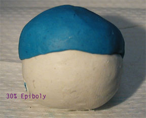
Figure 1. EVL (enveloping layer) at 30% epiboly (blue). White signifies yolk cells. |
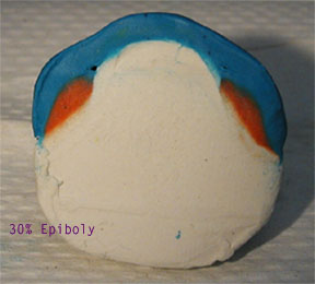
Figure 2. In a cross-section view (AP axis has been bisected), mesoderm (red) is visible, as is the dome of the EVL over the deep embryonic cells. |
 |
 |
|
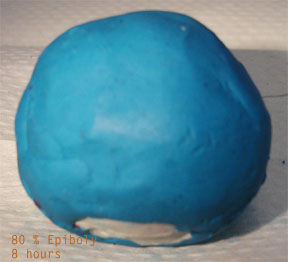
Figure 3. At 8 hours, 80% epiboly of the EVL is seen. At the remaining bottom opening, yolk can be seen. |
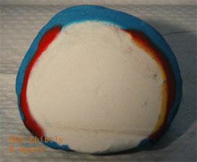
Figure 4. On the left, mesodermal precursors (red) can be seen to have migrated downwards. On the right, mesendodermal cells (orange) line the ectoderm precursor (blue). The ectoderm gives rise to skin and neural tissue, whereas the mesoderm gives rise to somites and mesenchymal cells. Endoderm will become internal organs. |
 |
 |
|
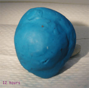
Figure 5. At 12 hours, epiboly is
complete. The raised notch of the notochord and presumptive
spinal chord can be seen.The embryo will form
asymmetrically, to one side of the yolk (see Figure 6).
|
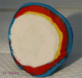
Figure 6. Again, in a cross-sectional view, the mesoderm underlies the ectoderm (ventral-dorsal axis is left to right in this photograph), which covers the endoderm (yellow). At the former vegetal pole, there is only yolk and a thin layer of ectoderm. |
 |
 |
|
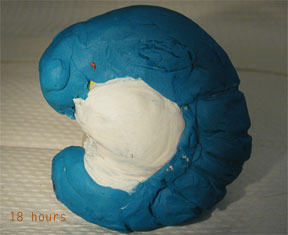
Figure 7. At 18 hours, gastrulation is complete. At this point, the embryo has eye, vertebrate, pharyngeal, and tail precursors. |
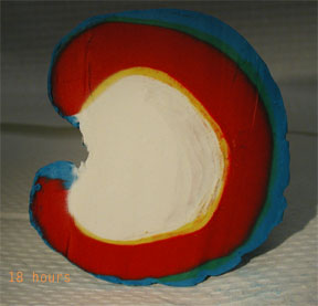
Figure 8. In a cross-sectional view, the embryo can be seen to occupy the right side of the mass. The yolk will shrink until the left side (ventral ectoderm) meets the yellow endoderm on the right side. |
Fascinated? Go to the Developmental Biology Website by Scott Gilbert to Learn More!
References:
1. Gilbert, Scott F., Developmental Biology, Sinauer Associates, Inc. Massachusetts, 2003. Textbook for the Swarthmore Developmental Biology Course
2. Gilbert, Scott F., Developmental Biology, www.devbio.com, 2003. Accompanying website for the textbook
3. Cebra Thomas, Judy, Biology 24 Lectures (personal communication), Swarthmore College, 2004.