|
Results:
The embryos of the first
experiment were treated with one of two growth factors,
either bFGF or VEGF. Those treated with bFGF showed the
greatest amount of blood vessel forma- tion when compared
with the DMEM and VEGF embryos (pictures
of the results). A problem may
have occurred with the VEGF, as it had been in the lab for
an extended period of time and may have been too old to
produce accurate results.
Of the
twelve embryos prepared for the second experiment (during
which only the effects of bFGF on blood vessel formation was
evaluated), seven survived. When opened, those embryos that
did not survive had significantly darker amnionic fluid than
their living counterparts. In addition, the amnionic fluid
of the dead embryos appeared cloudy and gave off a distinct
odor. Of the seven surviving embryos, only the filter paper
disks of three were removed successfuly (with portions of
the CAM membrane attached). In other cases, when removing
the disks, blood vessels were cut in such a way as to
contaminate the filter paper squares; some removed squares
were dyed red by the ruptured blood vessels, and the CAM
membrane was either dislodged or lost.
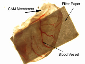
Figure
1. Photograph of CAM membrane attached to a filter paper
square (3mm x 3mm) treated
with DMEM. The CAM membrane was removed from a 15-day-old
chick embry, after the
membrane was exposed to a treated filter paper square for
five days.
When
examining the numbers of blood vessels on the CAM membranes
treated with either DMEM or bFGF, the one treated with bFGF
had a greater number of blood vessels (13) as compared with
the vessels observed on the two disks treated with DMEM (7
and 12, respectively); however, it must be taken into
account that only three test results were collected. In
addition, a significant difference between the number of
blood vessels of either the DMEM or bFGF CAM membranes did
not exist.
|